Leave message
Can’t find what you’re looking for?
Fill out this form to inquire about our custom protein services!
Inquire about our Custom Services >>


 Request a FREE Sample of our FcRn Binding Kit!
 Request a FREE Sample of our Fc gamma RI / CD64 Binding Kit !
Happy Holiday! Limited Keychain here with your next orderHappy Holiday! Limited Keychain here with your next order
Request a FREE sample of our GMP products! Request a FREE sample of our GMP products!
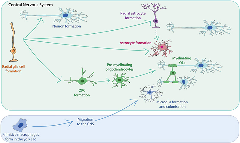
> Neural Antibodies | The Key to Precise Detection of Nerve Cell Markers 
| Molecule | Cat. No. | Product Description | Application |
|---|---|---|---|
| GFAP | GFP-S453 | Polyclonal GFAP Antibody, Rabbit IgG | Specific binding with GFAP to identify astrocytes |
| NG2/Cspg4 | NG4-S455 | Polyclonal NG2/Cspg4 Antibody, Rabbit IgG | Specific binding with NG2 to identify oligodendrocyte progenitors |
| Olig2 | OL2-S456 | Polyclonal Olig2 Antibody, Rabbit IgG | Specific binding with Olig2 to identify oligodendrocytes |
| Tbr1 | TB1-S457 | Polyclonal Tbr1 Antibody, Rabbit IgG | Specific binding with Tbr1 to identify immature neurons |
| NeuN/Rbfox3 | NE3-S454 | Monoclonal NeuN/Rbfox3 Antibody, Mouse IgG1 | Specific binding with NeuN to identify mature neurons |
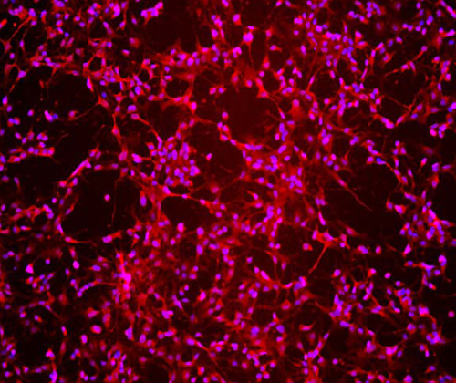
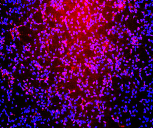
Immunofluorescent staining (10X) of cerebral organoid-derived cells (Cat. No. CIPO-BWL001K) labeling Tbr1 (Red) with purified Polyclonal Tbr1 Antibody, Rabbit IgG (Cat. No. TB1-S457) at 1:200 dilution. DAPI (blue) was used as nuclear counterstain.
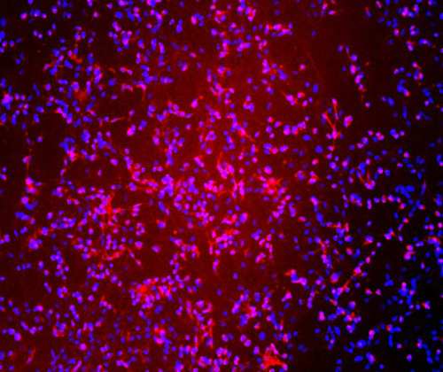
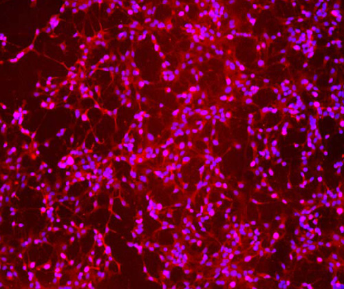
Immunofluorescent staining (10X) of cerebral organoid-derived cells (Cat. No. CIPO-BWL001K) labeling NeuN (Red) with purified Monoclonal NeuN/Rbfox3 Antibody, Mouse IgG1 (Cat. No. NE3-S454) at 1:500 dilution. DAPI (blue) was used as nuclear counterstain.
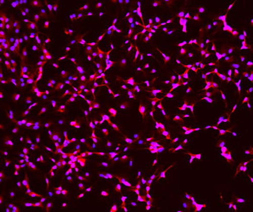
Immunofluorescent staining (10X) of cerebral organoid-derived cells (Cat. No. CIPO-BWL001K) labeling GFAP (Red) with purified Polyclonal GFAP Antibody, Rabbit IgG (Cat. No. GFP-S453) at 1:200 dilution. DAPI (blue) was used as nuclear counterstain.
Immunofluorescent staining (10X) of cerebral organoid-derived cells (Cat. No. CIPO-BWL001K) labeling NG2 (Red) with purified Polyclonal NG2/Cspg4 Antibody, Rabbit IgG (Cat. No. NG4-S455) at 1:200 dilution. DAPI (blue) was used as nuclear counterstain.
Immunofluorescent staining (10X) of cerebral organoid-derived cells (Cat. No. CIPO-BWL001K) labeling Olig2 (Red) with purified Polyclonal Olig2 Antibody, Rabbit IgG (Cat. No. OL2-S456) at 1:200 dilution. DAPI (blue) was used as nuclear counterstain.
1. Neely S A, Lyons D A. Insights into central nervous system glial cell formation and function from zebrafish[J]. Frontiers in Cell and Developmental Biology, 2021, 9: 754606. https://doi.org/10.3389/fcell.2021.754606
2. Yokoo H, Nobusawa S, Takebayashi H, et al. Anti-human Olig2 antibody as a useful immunohistochemical marker of normal oligodendrocytes and gliomas[J]. The American journal of pathology, 2004, 164(5): 1717-1725. https://doi.org/10.1016/S0002-9440(10)63730-3
3. Zhao W, Dumanis S B, Tamboli I Y, et al. Human APOE genotype affects intraneuronal Aβ1–42 accumulation in a lentiviral gene transfer model[J]. Human molecular genetics, 2014, 23(5): 1365-1375. https://doi.org/10.1093/hmg/ddt525
4. Yu L, Chen C, Wang L F, et al. Neuroprotective effect of kaempferol glycosides against brain injury and neuroinflammation by inhibiting the activation of NF-κB and STAT3 in transient focal stroke[J]. PloS one, 2013, 8(2): e55839. https://doi.org/10.1371/journal.pone.0055839
5. Rigo Y R, Benvenutti R, Portela L V, et al. Neurogenic potential of NG2 in neurotrauma: a systematic review[J]. Neural Regeneration Research, 2024, 19(12): 2673-2683. https://doi.org/10.4103/NRR.NRR-D-23-01031
6. Englund C, Fink A, Lau C, et al. Pax6, Tbr2, and Tbr1 are expressed sequentially by radial glia, intermediate progenitor cells, and postmitotic neurons in develo** neocortex[J]. Journal of Neuroscience, 2005, 25(1): 247-251. https://doi.org/10.1523/JNEUROSCI.2899-04.2005
7. Jurga A M, Paleczna M, Kadluczka J, et al. Beyond the GFAP-astrocyte protein markers in the brain[J]. Biomolecules, 2021, 11(9): 1361. https://doi.org/10.3390/biom11091361
8. Alekseeva O S, Gusel’nikova V V, Beznin G V, et al. Prospects for the application of neun nuclear protein as a marker of the functional state of nerve cells in vertebrates[J]. Journal of Evolutionary Biochemistry and Physiology, 2015, 51: 357-369. https://doi.org/10.1134/S0022093015050014


This web search service is supported by Google Inc.


 A-Z
A-Z